

What to Know About: Plantar Fasciitis (For Providers)
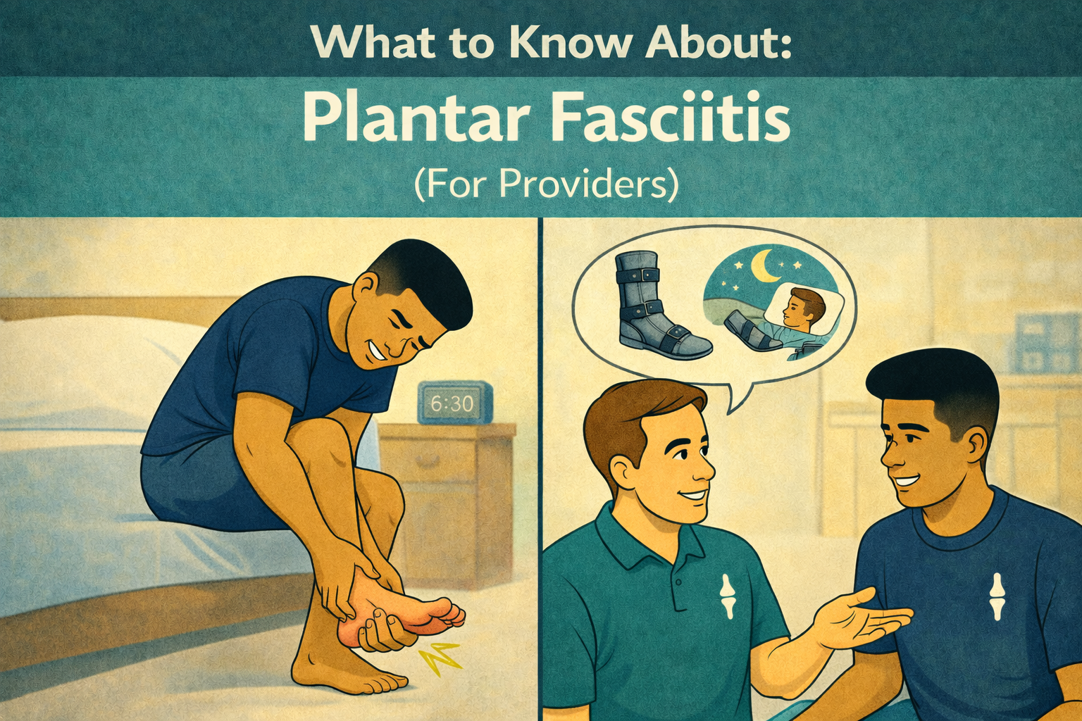
If you’ve ever stepped out of bed and felt a sharp pain in your heel that made you pause and brace yourself—you’re not imagining it. That “first-step pain” is one of the most classic signs of plantar fasciitis, one of the most common foot conditions adults experience.
But plantar fasciitis is rarely just about the heel.
It’s about how your ankle moves, how your toes function, how your arch supports you, and how all of that connects to the way you walk, work, and live.
Let’s break it down.
✨ Too Long Didn’t Read (TL;DR) / Summary
Plantar fasciitis is irritation or degeneration of the plantar fascia on the bottom of the foot.
Pain is often worst with the first steps in the morning or after long-term sitting.
It’s influenced by ankle stiffness, calf tightness, foot strength, toe mobility, and arch mechanics.
High arches and low arches can both contribute—there’s no single “bad foot.”
Most people (>80%) improve with conservative care, especially physical therapy.
Manual therapy, calf stretching, foot strengthening, and load management matter.
🧾 Condition-Specific General Information
What is plantar fasciitis?
Plantar fasciitis is a condition involving the plantar fascia, a thick band of connective tissue that runs from the heel to the toes and helps support the arch of the foot.¹²
Despite the “-itis” in the name, research shows that many cases—especially chronic ones—reflect degeneration rather than inflammation. Because of this, some experts also use the term plantar fasciosis or plantar fasciopathy.
👉 Translation for patients:
This tissue gets overloaded faster than it can recover.
Common symptoms patients report
Heel or arch pain with first steps in the morning
Pain after sitting, then standing up
Pain that improves with movement—but returns later
Tenderness at the inside of the heel
Rarely: swelling (bruising may suggest a different injury)
Why the ankle matters (more than most people realize)
Limited ankle dorsiflexion—often due to gastroc or soleus tightness—is one of the most consistently reported risk factors for plantar fasciitis.
When the ankle can’t move well:
The foot compensates
The arch absorbs more stress
The plantar fascia takes the hit
This is why calf stretching isn’t “basic” care—it’s foundational care.
High arch vs. low arch: which is worse?
Neither. Both. Research is mixed but says there’s no “better” option.
High arches may increase tensile load on the plantar fascia.
Low or pronated arches may increase strain due to prolonged loading.
What matters more than arch type is how the foot adapts under load.
The big toe, the arch, and the “windlass”
The great toe (aka: “the hallux”) plays a critical role in plantar fascia tension through the windlass mechanism.
Too little toe motion (hallux limitus)
Structural changes (hallux valgus)
Poor coordination during gait
All can increase plantar fascia strain.
Side Note: The windlass mechanism is the way your big toe, arch, and plantar fascia work together to support your foot during walking and standing. When your big toe bends upward as you push off the ground, it tightens the plantar fascia—much like winding a rope around a winch—which lifts and stiffens the arch to make your foot more stable. If the big toe or foot can’t move well, that system doesn’t work as smoothly, and extra strain can show up as foot or heel pain.
Foot intrinsic muscles: the quiet stabilizers
Smaller muscles within the foot help:
Support the arch
Control toe motion
Reduce strain on passive structures like the plantar fascia
Weakness here has been associated with plantar fasciitis and foot pain. Strength training for these muscles—done correctly—can reduce pain and improve function.
Other known risk factors
Increased BMI
Prolonged standing or repetitive loading
Sudden increases in walking or running volume
Decreased ankle mobility
Inadequate recovery time
Age (most common between 40–60 years)
👩⚕️ For Providers 👨⚕️
The conversation patients need (but don’t always get)
Many patients arrive believing:
“I just need better shoes.”
“I must have bad arches.”
“I should stop moving.”
Reframing matters. Providers must start their care with good education.
Helpful language:
“This tissue is overloaded—not broken.”
“We’re going to improve how your foot and ankle share the load.”
“Pain doesn’t mean damage—it means we need a smarter plan.”
Diagnosis: less imaging, more listening
Clinical history and physical exam remain the cornerstone of diagnosis. Routine imaging typically is not required unless:
Symptoms persist
The history is unclear
Another diagnosis is suspected
Ultrasound can be useful for evaluating thickness and guiding treatment, but it should support—not replace—clinical reasoning.
Evidence-based physical therapy interventions
Current guidelines and reviews support:
Manual Therapy (A-Level Evidence)
Soft tissue and joint mobilization (including talocrural joint)
Stretching (A-Level Evidence)
Gastrocnemius and soleus
Plantar fascia–specific stretching
Strengthening (B-Level Evidence)
Foot intrinsics
Ankle musculature
Progressive loading when tolerated
Night Splints (A-Level Evidence)
1–3 months for patients with morning pain
Taping (A-Level Evidence)
Short-term symptom relief
Dry Needling (B-Level Evidence)
Evidence supports short- and long-term pain reduction
Modalities
Therapeutic ultrasound is not recommended
Low-level laser therapy may help short-term pain
Iontophoresis can be considered as a secondary option
What about injections and shockwave?
Corticosteroid injections may help short-term pain but vary widely in outcomes
PRP may show promise for longer-term relief in chronic cases but more data is needed
Extracorporeal shockwave therapy has mixed but growing evidence
These options work best when paired with targeted therapeutic rehabilitation and movement, not used in isolation.
Surgery: the exception, not the rule
Surgery is reserved for a small subset who fail extensive conservative care. Endoscopic procedures generally allow faster return to work and fewer complications compared to open techniques but, oftentimes, plantar fasciitis does not require surgery.
Key Takeaways
Morning heel pain is common—but not something you have to “push through.”
Shoes help—but they don’t replace strength and mobility.
Stretching, strengthening, and load management work best together.
Most people improve without surgery.
The best outcomes happen when patients and providers build a plan together.
📂 Supplemental Information / Citations
Tseng WC, Chen YC, Lee TM, Chen WS. Plantar fasciitis: An updated review. J Med Ultrasound. 2023;31(4):268-274.
Motley T. Plantar fasciitis/fasciosis. Clin Podiatr Med Surg. 2021;38(2):193-200.
Koc TA Jr, Bise CG, Neville C, et al. Heel pain—plantar fasciitis: Revision 2023. J Orthop Sports Phys Ther.2023;53(12):CPG1-CPG39.
Huffer D, Hing W, Newton R, Clair M. Strength training for plantar fasciitis and the intrinsic foot musculature. Phys Ther Sport. 2017;24:44-52.
Voelker R. What is plantar fasciitis? JAMA. 2024;332(13):1120.
Aranda Y, Munuera PV. Plantar fasciitis and its relationship with hallux limitus. J Am Podiatr Med Assoc.2014;104(3):263-268.
Cobden A, Camurcu Y, Sofu H, et al. Evaluation of the association between plantar fasciitis and hallux valgus. J Am Podiatr Med Assoc. 2020;110(2).
This content drafted, researched, edited, and generated by:
Jackson Kojima, PT, DPT
Jackson Kojima, PT, DPT, OCS is a physical therapist with an extensive background in orthopedics, geriatrics, and sports rehabilitation. Dr. Kojima is a board-certified orthopedic clinical specialist (OCS) with a passion for post-operative rehabilitation and enjoys treating multi-factorial conditions like low back pain and generalized joint pain. Dr. Kojima earned his doctorate of physical therapy from Campbell University in 2021 and currently practices in Greenville, SC.
© 2026 The Joint Connection Company. All rights reserved.
The content on this website, including all text, graphics, and materials, is the exclusive property of The Joint Connection Company and is protected by applicable copyright and intellectual property laws. No part of this site may be reproduced, distributed, or used without prior written permission.